

Need trusted radiology services in Delhi NCR? Maccure Hospital offers expert care with a team of leading radiologists dedicated to precise diagnosis and effective treatment. Using advanced technology and a personalised approach, Maccure ensures accurate imaging and reliable results, supporting every patient’s health journey with excellence.

From your initial consultation to your final diagnosis, our dedicated team of radiologists in Delhi NCR is here to guide and support you every step of the way.
At Maccure Hospital, we offer a comprehensive range of radiology services to meet diverse medical needs. Here are some of our services:
1. Early Pregnancy
An early pregnancy scan, also known as a viability or dating scan, helps confirm pregnancy, estimate the due date, and assess the health of the baby. Conducted between the 7th and 12th week, this scan provides a clear view of the pregnancy sac, number of embryos, and heartbeat, ensuring the pregnancy is progressing well. Doctors may perform the scan abdominally or transvaginally, depending on clarity.
2. Foetal Echo
Foetal echocardiography is a specialised ultrasound to examine the baby’s heart structure and function. Done between 18 and 24 weeks, this scan is advised if there is a family history of congenital heart issues, genetic abnormalities, or other maternal conditions. The test helps identify and monitor any heart defects early, ensuring proper care.
3. Anomalies Detection
The anomaly scan, also known as the mid-pregnancy scan, is performed between 18 and 21 weeks to check for physical abnormalities. During the scan, doctors examine the baby's bones, brain, heart, and other organs. This scan detects conditions like cleft lip, spina bifida, and other serious abnormalities, ensuring parents are well-informed.
4. Interval Growth
An interval growth scan, usually done in the third trimester, assesses the baby’s growth and position, the amount of amniotic fluid, and the position of the placenta. Recommended for mothers with gestational diabetes, high blood pressure, or other pregnancy concerns, this scan helps doctors monitor the baby’s health and prepare for delivery.
5. Paediatric Ultrasound
A paediatric ultrasound is a safe, non-invasive examination of a child’s abdomen. This test uses sound waves to produce images of organs like the liver, kidneys, and intestines. It is especially helpful for diagnosing abdominal pain or conditions in young patients, as it provides accurate imaging without radiation.
6. X-Ray
An X-ray is a widely used imaging technique that uses low radiation levels to capture detailed images of internal structures, such as bones and organs. It assists doctors in diagnosing fractures, infections, and other internal issues, making it an essential tool in medical diagnosis and treatment.
Fill this form to get instant support by our expert team.

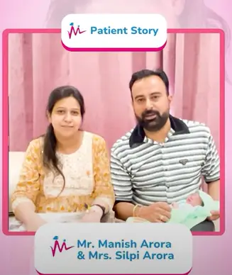
We know that starting or growing a family can be filled with challenges, both emotional and physical. And so, we strive to provide not only exceptional medical care but also a warm, supportive space where families can come together during this exciting time.

1 year ago
We had a three days stay at Maccure Hospital during the delivery of our baby boy. I have to say that everything about the hospital is just outstanding from its infrastructure to the staff. All the staff including doctors, nurses, housekeeping and the hospital staff were really caring, loving and hospitable. We will really recommend Maccure to everyone who want a great experience while being their babies into this world. Thanks to everyone at Maccure Hospital.

1 year ago
Nice experience doctor and staff , facilities top notch, Clean and comfortable,and highly recommended

1 year ago
Dr. Sanjay Jain is not only an excellent doctor, he is a simple, sober and super human being. He is very friendly, approachable with always a smiling face. He is very intelligent, dedicated and one of the most experienced doctors I have ever met. He impressed me with his simple personality I got relieved from my stress in the very first meeting with him. He has not only become my good friend but also I give him place of God in my life. All the very best and my good wishes to him always.

1 year ago
My wife delivery was done 15 days before at Maccure Hospital, Janakpuri. Bringing our baby into the world at this hospital was an unforgettable experience. The maternity team was incredibly supportive, guiding us every step of the way with warmth and expertise. From prenatal care to delivery, we felt confident and well-cared-for. Grateful for the exceptional staff who made our journey to parenthood so special.The loveliest part was the decoration photoshoot and cake cutting celebration 😊👍

1 year ago
Exceptional care at Maccure Hospital! JanakpuriThe staff is compassionate, efficient, and truly dedicated to patient well-being.From the moment we walked in, the atmosphere was welcoming.The doctors and nurses provided top-notch service, ensuring a smooth and comfortable experience. Grateful for their commitment to healthcare excellence

2 years ago
The doctor and the nursing staff were excellent in taking care of the baby and the mother during the time in hospital. Dr S.K.Jain was very thorough with his explanations and was patient while answering our queries. Shout out to Dr Geeta, nurse Mukeema and nurse Priyanka for their care and support during our stay. The entire team was very patient and the baby was delivered normally after almost 12 hours.Kudos to the NEW "MACCURE".

8 months ago
Dr Geeta Jain mam very good experienced Doctor in Delhi NCR
A1. We provide early pregnancy scans, foetal echocardiography, anomaly detection, interval growth scans, paediatric ultrasounds, X-rays, and more to meet diverse diagnostic needs.
A2. Yes, most scans such as ultrasounds are completely safe during pregnancy, as they use sound waves instead of radiation. X-rays are avoided unless absolutely necessary.
A3. Paediatric ultrasounds help diagnose issues in children such as abdominal pain, organ abnormalities, and kidney or liver concerns without using radiation.
A4. An anomaly scan is best performed between 18 and 21 weeks of pregnancy to detect any physical abnormalities in the baby\u2019s organs and structures.
A5. Interval growth scans, typically conducted in the third trimester, assess the baby\u2019s growth, position, placenta health, and amniotic fluid levels to ensure optimal delivery planning.
A6. X-rays involve minimal radiation and are safe when used appropriately. They are commonly required to diagnose fractures, infections, or chest abnormalities.
A7. Foetal echocardiography focuses on examining the baby\u2019s heart structure and function. It is recommended between 18 and 24 weeks, especially if there\u2019s a family history of heart issues.
A8. Advanced radiology tools at Maccure Hospital enable early detection of abnormalities, supporting timely interventions and better outcomes for patients.
A9. Preparation depends on the type of scan. For example, a full bladder may be required for some pregnancy scans. Our team will provide detailed instructions during appointment confirmation.
A10. Maccure Hospital offers cutting-edge technology, experienced radiologists, and personalised care, ensuring accurate diagnoses and a compassionate experience for all patients.
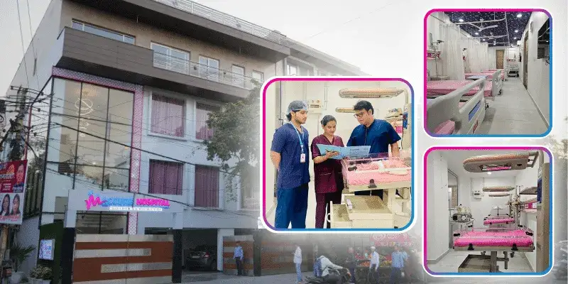
If you're seeking the perfect place to welcome your little one, Maccure Hospital stands as one of the top maternity & child care hospitals in Delhi NCR. We provide comprehensive maternity and paediatric care for you and your baby, ensuring a joyful experience.